Cone Beam CT (CBCT) in Orthodontics: The 3D X-Ray That Changes Everything

What Is Cone Beam CT (CBCT)?
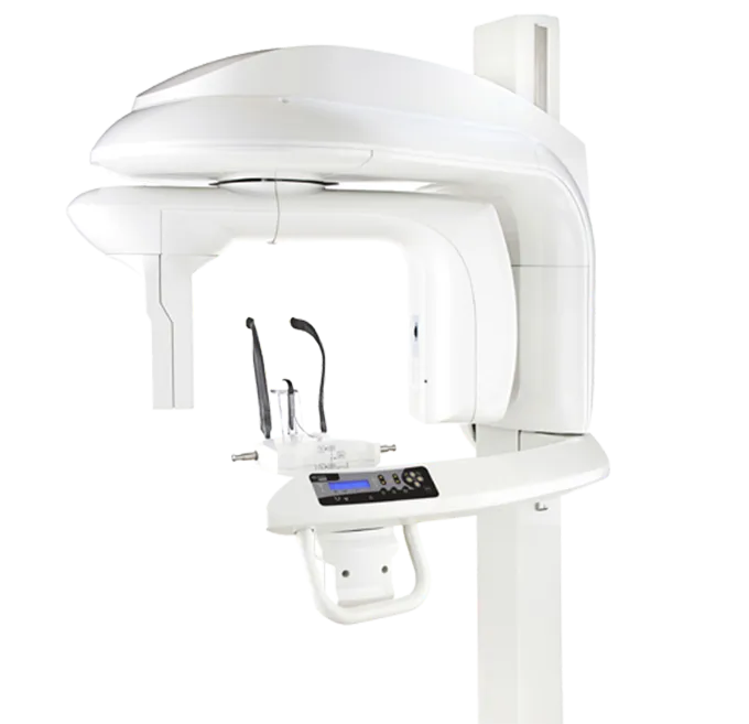
Cone Beam CT — also known as cone beam computed tomography or simply 3D X-ray — is an imaging technology that has transformed the way we diagnose and plan orthodontic treatments. Unlike a traditional panoramic X-ray that produces a flat two-dimensional image, the CBCT generates a complete three-dimensional model of your teeth, jaws, temporomandibular joints, and even your airways.
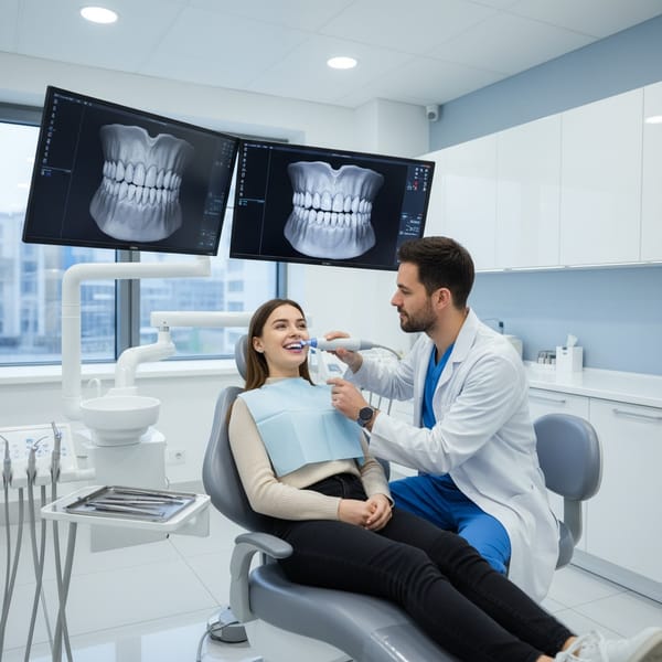
At the Rive Nord Orthodontic Centre, we have integrated this technology directly into our St-Eustache clinic. This means the examination is done on-site, in just seconds, without having to travel to an external radiology centre.
How Does CBCT Work?
The principle is elegant in its simplicity. A cone-shaped X-ray beam performs a single 360-degree rotation around your head. In 10 to 20 seconds, the machine captures hundreds of images from different angles. Sophisticated software then reconstructs these images into a 3D model that we can explore from every angle, zoom into, measure, and even use to simulate tooth movements.
It's a bit like going from Google Maps in satellite view (2D) to Google Earth in 3D: you suddenly see structures in depth, the relationships between elements, and details hidden behind overlapping layers.
CBCT vs. Panoramic X-Ray: What's the Difference?
The panoramic X-ray remains an essential tool in orthodontics. It provides an overall view of the dentition, roots, and surrounding structures. However, it has important limitations:
| Criteria | Panoramic (2D) | CBCT (3D) |
|---|---|---|
| Type of image | Flat, flattened image | 3D volume explorable in all planes |
| Overlapping | Structures overlap each other | No overlapping — each structure can be isolated |
| Measurement accuracy | 15-25% distortion | Submillimetre precision (0.1 to 0.4 mm) |
| Root visualization | Limited by overlapping | Each root visible individually |
| Airways | Cannot be assessed | Complete volumetric analysis |
| Joint (TMJ) | Summary view | Detailed 3D assessment |
| Exam duration | ~15 seconds | 10-20 seconds |
In summary, the CBCT does not systematically replace the panoramic, but it provides an additional dimension where 2D is no longer sufficient.
Applications of CBCT in Orthodontics
In my daily practice, CBCT proves indispensable in several clinical situations:
1. Impacted or Ectopic Teeth
Impacted canines are one of the classic cases where CBCT changes everything. In 2D, it is often difficult to determine the exact position of a tooth that hasn't erupted: is it on the palatal side? The labial side? Is it touching the root of a neighbouring tooth? CBCT answers all these questions with submillimetre precision, allowing us to plan orthodontic traction optimally and minimize the risk of damage to adjacent teeth.
2. Airway Evaluation
For patients suffering from sleep apnea or respiratory disorders, CBCT allows us to measure the volume and morphology of the upper airways in three dimensions. This information is essential to determine whether a mandibular advancement device could improve breathing, or whether a surgical approach is necessary.
3. Temporomandibular Joint (TMJ) Disorders
Jaw pain, clicking, and limited opening are often related to joint abnormalities. CBCT allows visualization of condyle shape, joint space, and any bone degeneration with precision impossible to achieve in 2D.
4. Orthodontic Mini-Screw (TAD) Planning
Mini-screws are small temporary anchors that we insert into the bone to achieve tooth movements otherwise impossible. CBCT allows us to measure available bone thickness, root positions, and sensitive anatomical structures for safe and precise placement.
5. Early Detection of Root Resorption
Root resorption — a progressive shortening of the roots — is sometimes a side effect of orthodontic treatment. CBCT detects these changes earlier and with greater sensitivity than conventional radiography, allowing us to adjust the treatment before the problem worsens.
6. Palatal Expansion and Asymmetries
For patients with a narrow palate, CBCT evaluates the thickness of the midpalatal suture and the amount of available bone. For patients with facial asymmetries, 3D imaging is the only way to truly understand the origin of the discrepancy — skeletal, dental, or both.
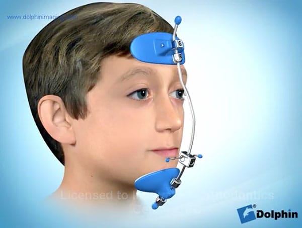
7. Orthognathic Surgery
For cases requiring jaw surgery, CBCT is indispensable. It allows the surgeon and the orthodontist to plan bone movements together in 3D, simulate the outcome, and fabricate custom surgical guides for a more precise and predictable procedure.
What About Radiation Dose?
This is a legitimate and important question. Here are the facts:
- A latest-generation CBCT emits a radiation dose significantly lower than a conventional medical CT scan (approximately 80% less)
- The dose is comparable to that of 4 to 8 panoramic X-rays, depending on the field of view used
- For reference, a round-trip flight from Montreal to Paris exposes you to approximately 0.1 mSv of cosmic radiation — equivalent to one or two CBCTs
We rigorously follow the ALARA principle (As Low As Reasonably Achievable): CBCT is only prescribed when the 3D information provides real diagnostic value that the 2D X-ray cannot deliver. It's not systematic — it's judicious.
The Patient Experience: Simple and Quick
If you've never had a CBCT, here's what to expect:
- You remain standing or seated (not lying down like in a medical CT scanner)
- You place your chin on a support
- The machine rotates around your head for 10 to 20 seconds
- That's it — no pain, no particular sensation
The images are immediately available on our screen, and I can show you your teeth, roots, and jaws from every angle in real time. Many patients find this step fascinating — it's a bit like seeing inside their own smile for the first time.
CBCT at the Service of Your Personalized Treatment
What fascinates me about this technology is the personalization it enables. Every patient is unique — jaw shape, bone thickness, root positions, airway volume — and CBCT gives us access to this individuality with remarkable precision.
Combined with our intraoral scanners (3D digital impressions) and digital treatment planning, CBCT is part of a technological ecosystem that allows us to:
- Make more precise diagnoses
- Plan more predictable treatments
- Reduce surprises during treatment
- Communicate more clearly with you about your care plan
When Is CBCT Recommended?
CBCT is not necessary for all patients. It is particularly useful in the following situations:
- Impacted teeth (canines, wisdom teeth) requiring precise localization
- Facial asymmetries or jaw discrepancies
- TMJ disorders with pain or dysfunction
- Sleep apnea evaluation
- Orthodontic mini-screw planning
- Cases requiring orthognathic surgery
- Monitoring root resorption
- Complex cases requiring in-depth analysis
During your consultation, I will determine whether a CBCT is relevant for your situation. If a panoramic X-ray is sufficient, that's what we'll use. The right technology at the right time — that's our approach.
Have Questions?
If you're curious about how CBCT could contribute to your orthodontic diagnosis, don't hesitate to ask during your next visit. At the Rive Nord Orthodontic Centre, we offer a free consultation for all new patients — the perfect opportunity to discover our technological capabilities and discuss your options.
Dr. David Benguira, Orthodontist
Rive Nord Orthodontic Centre, St-Eustache